Managing pain in post total knee replacement plays a key role in successful outcomes
Osteoarthritis (OA) affects approximately one in 10 individuals and is equally prevalent in men and women, although there is higher prevalence in postmenopausal and obese women.1,2,3 Knee OA’s high prevalence is directly associated with a rising number of surgical interventions designed to reduce pain and increase mobility for patients (ie, total knee arthroplasty [TKA]).
Per Moffet et al’s 2004 study, “More intensive rehabilitation should be promoted in the subacute recovery period after TKA, to optimize functional outcomes in the first year after surgery.”4 Aggressive focus on active and passive knee extension is one of the most important components of postoperative early rehabilitation. Persistent TKA flexion contractures are difficult to eliminate once they have been present for more than 3 months.5
The most prominent outcome variable linked to OA and TKA is pain.1,6,7,8 The process for pain management that was applied in prehabilitation is the same process and rationale for postsurgical pain management.
Central sensitization plays an important role in postsurgical and post-traumatic pain.9,10 Postoperative pain is mostly nociceptive, which is pain perception following surgical insult.
Case Study
Patient P. Tella, male, aged 76 years old, presented post left TKA June 2, 2014, with multiple comorbidities. Postop at evaluation, P. Tella presented with postoperative pain, swelling, and bruising (typical of the recovery process following knee surgery). He also demonstrated muscle weakness, as would be expected with any surgery. All the cardinal signs of inflammation were present at the knee and its periphery. Different functional reporting tools (Functional Gait, PANAS, Pain Disability Index, Lower Extremity Functional Scale) were used at evaluation, at each visit, and at the discharge to determine efficacy of the interventions and to gauge progress at any stage of treatment.
A constant passive motion (CPM) machine11, such as those available through The Furniss Corporation, Grove City, Ohio, or the Optiflex by Patterson Medical, Warrenville, Ill, was used immediately postsurgery and continued at home x 2 weeks, 2 hours, three times a day (TID).
Bruising caused additional tenderness. As in prehabilitation, inflammation management will reduce pain. Each visit’s treatment included the use of photo-biostimulation over terminal sites in the inguinal region to open up the lymphatic flow to decrease inflammation from distal to proximal. Chattanooga, Tenn-based Richmar’s MedX 785-nm laser with 200 mW Gallium-Aluminum-Arsenide diodes on the EVO CM4 provided quick treatment of the terminal sites at 4 joules/cm2 or 20 seconds (the laser generates 1 joule every 5 seconds). This included use of superluminous diodes (SLD) at the terminal site post laser to coincidentally treat the terminal sites while applying ultrasound with the same Richmar EVO CM4 to reduce inflammation at the local site. The SLD infrared (870 nm) and the visible red (633 nm) provide similar outcomes to the laser to open up those terminal sites, but takes a little longer at 1 Joule per 45-second application (or a 3-minute treatment) to be applied every other day. SLD was applied each visit with the same parameters over the terminal sites. The market also includes Class IV lasers from Irvine, Calif-based Zimmer MedizinSystems, such as the OptonPro Class IV laser, which was recently released into the United States. Other devices include the Apollo Class IV cold laser from PHS Therapeutics, a division of Pivotal Health Solutions, Watertown, SD, and MR4 Super Pulsed Laser, Multi Raidance Medical, Solon, Ohio.
Coincidentally with photo-biostimulation, the 3 MHz sound head at 0.5 w/cm2 x 10 minutes (5-minute treatment for two times the size of the soundhead or, more specifically, the Effective Radiating Area for that soundhead) was used over the bruised and inflamed edematous, which will change cell membrane permeability by a process of acoustic streaming and stable cavitation. Richmar’s EVO CM4 ultrasound cued up the subthermal application with a single button using the hands-free Accusound applicator. Ultrasound will also modulate vasoconstriction; change lymphocyte adhesion properties of the endothelium; increase mast cell degranulation; increase phagocytosis by macrophage; increase production of growth factors by macrophages, resulting in calcium fluxes in fibroblasts, angiogenesis, proliferation of T-cells, osteoblasts, fibroblasts, and a number of proteins associated with inflammation and repair (IL-1, IL-2, IL-6, IL-8, interferon-?, fibroblast growth factor-b, vascular endothelial growth factor, collagen),12-25 as well as accelerate thrombolysis26-35; or reduce inflammation.
Use of the Richmar EVO CM4 will activate the appropriate parameters for the muscle pump using the following parameters: Russian Stimulation current, ramp time up 2 seconds, ramp time down 2 seconds, on time of 4 seconds and off time of 12 seconds, and treatment time of 10 minutes. With the limb elevated, electrodes are placed on the muscles on the quadriceps and the hamstrings with the intensity increased to a vigorous muscle twitch to wick away the inflammation to the aforementioned open terminal sites. All this is done coincidentally (biostimulation, ultrasound, and e-stim), saving time and increasing the quality of outcomes.
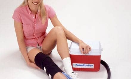
Fan cut therapeutic taping technique using Kinesio Tape from Kinesio Holding Corp, Albuquerque, NM, was applied with the base of the tape at the inguinal terminal site, and no tension along the length of the tape from the terminal site to surrounding the knee was utilized each visit to allow a passive means to carryover inflammation or edema management done in the clinic. P. Tella was instructed to elevate the lower extremity higher than heart level for an hour or two BID incorporating ankle pumps to manage the edema at home. The use of ice packs, such as those available through Battle Creek Equipment, Fremont, Ind, or cold packs by Strongsville, Ohio-based Roscoe Medical, can offer supplemental pain control through cold therapy, restricting the blood vessels and slowing the nerve impulse. To this end, ice pack use three to four times a day for about 10 to 20 minutes was instructed to reduce swelling and inflammation at the knee joint and surrounding tissue.
Per the pain modulatory mechanism of the “Gate Control” theory proposed by Melzack and Wall in 196536 and Bausbaum and Fields’37 proposed endogenous opiate theory, pain management was achieved with electrical stimulation (e-stim). Different frequencies activate central mechanisms to produce analgesia. Low-frequency e-stim activates ?-opioid receptors in the spinal cord and the brainstem, whereas high-frequency e-stim activates opioid receptors in the spinal cord and the brainstem.38-40 Any e-stim device, whether a clinic-based unit or any portable TENS unit such as the Richmar EVO CM4, the BioStim SD digital TENS, BioMedical Life Systems Inc, Vista, Calif, or SpectrumMicro-100, from Amrex-Zetron Inc, Paramount, Calif, would use the gate control or the endogenous opiate theory to result in similar outcomes, even though wave forms are different.
Photo-biostimulation with similar parameters as for inflammation management can be used but over acupuncture points in the arm at LI4, LI1, Tb5, and P6 as an effective means of managing pain anywhere in the body with the doses mentioned above. However, at each visit with P. Tella, the preset button on the Richmar EVO CM4 cued up Interferential current (IFC) quadpolar electrode placement (chained 100 Hz x 15 minutes and 2 Hz x 15 minutes, vector on fast 90o with an intensity at a slight motor twitch was utilized for pain management. Microcurrent (chained 30 Hz x 15 minutes and 0.3 Hz x 15 minutes with a subsensory intensity) could be utilized if the patient did not tolerate the sensation of IFC e-stim. Electrodes were applied to the same four acupuncture points at LI4, LI11, Tb5, and P6 to affect Melzack and Wall’s Gate Control Theory41 or Bausbaum and Fields42 endogenous opiate theory. A portable TENS unit with the same 100 hHz x 30 minutes and 2 pps x 30 minutes and a pulse width of 300 µsec was instructed to allow the means to carry over the positive pain-management results at home.42 He was instructed to use the TENS as often as he had pain to improve and maintain his pain threshold. Hyperalgesia or pain relief occurs whether an asymmetrical or symmetrical waveform is used. Different waveforms can be used to improve comfort but not to increase analgesic efficacy43, hence the efficacy of the TENS for home use.
Topical Creams and Patches
Topical creams and patches (first-generation transdermal delivery systems44) applied to the knee may help reduce pain. These products usually include active ingredients like capsaicin (also found in chili peppers), menthol, or salicylates. When absorbed through the skin, these ingredients are known to ease pain. Moab, Utah-based Sore No More, features the active ingredient menthol in its topical analgesic, Sore No More, which targets arthritis pain relief. Similar products for minor aches and pains are Flexall gels from Ari-Med Pharmaceuticals, Tempe, Ariz, and Sombra Professional Therapy Products, Albuquerque, NM, which offers pain-relieving gels comprised of warming and cool ingredients.
LidoFlex patches (nonprescription) were used at the periphery of the patella and changed out every 12 hours. P. Tella used the patches during waking hours at home as part of his pain-management regimen. Passive diffusion across the skin45-47 with transdermal patches eliminate frequent dosing administration and plasma level peaks and valleys associated with oral dosing and injections to maintain a constant drug concentration, and a drug with a short half-life can be delivered easily.
Second-generation transdermal delivery systems44 include those in which a substance bearing a charge is propelled through the skin by a low electrical current. Iontophoresis is defined as a means of use of a continuous direct electrical current to deliver therapeutically charged ions across the skin and into the systemic circulation.48-51 The IontoPatch, Patterson Medical, Warrenville, Ill, and the ActivaPatch, available through Gilroy, Calif-based ActivaTek Inc, exemplify technologies used in the delivery of iontophoresis that are available on the market. Phonophoresis would also be alternative to enhance the absorption of topically applied analgesics and anti-inflammatory agents through the therapeutic application of ultrasound. Neither of these techniques were used with P. Tella. Ultrasound applicator gels can be found through companies such as Parker Laboratories, Fairfield, NJ, which offers Aquasonic Clear and SCAN ultrasound gel.
Gaining strength in the muscles that support the joint in prehabilitation is as much of a requirement in post surgical management to help support the stability of the knee joint.
The reverse order of motor unit recruitment combined with external loading (muscle hypertrophy) imposed on stimulated muscles during training is likely responsible for the gains in muscular strength observed during muscle re-education training. Studies suggest 30% more force than maximum voluntary contraction.52 Studies also suggest that e-stim for muscle re-education produces lasting gains in muscle force through a short-term training regimen (approximately 30 training sessions).36,53-56
P. Tella was provided and instructed in the use of a portable combination TENS/Electrical Muscle Stimulator (Twin Stim II) unit with similar parameters completing the carryover of muscle re-education on the days he was not in the clinic. Kinesio tape application I strip six squares in length was applied from origin to insertion with 50% tension to facilitate the hamstrings and the quadriceps musculature at each visit (protocol was for 5 days of use, 1 day rest, and reapplication on the seventh day).
Altered Gait Biomechanics and Pain
P. Tella demonstrated altered gait biomechanics of reduced walking speed/cadence and decreased motion57 and increased lateral trunk lean58all of which can be a result of pain and inflammation. Tempe, Ariz-based Mobility Research’s LiteGait was utilized at each visit to provide better posture, better balance and weight bearing assistance while correcting upright postures appropriate for ambulation both over a treadmill and subsequent carryover to over ground ambulation. The evidence across animal and human literature suggests the number of repetitions for cortical reorganization through neuroplasticity is in the hundreds for the upper extremity59 and thousands for gait steps60 and with the LiteGait, P.Tella was able to attain 3168 feet first visit and by last visit 5280 feet. LiteGait enabled a partial weight bearing environment to allow the freedom for the therapist to facilitate weight shifting to correct P.Tella’s antalgic gait by off loading sufficiently the lower extremity associated with the painful post surgical knee initially. This allowed facilitation of foot placement and cueing appropriate patterns of walking. LiteGait through the support straps allowed progressive increase in weight bearing successively through each visit allowed distribution of supporting forces to meet P.Tella’s needs to become more symmetric in static posture and more functional in dynamic gait. LiteGait allowed an increased stretch of the hip flexors to facilitate the central pattern generators (automatic gait) and to integrate knee extension to increase painfree weight bearing in mid stance (often negated with lower extremity pathologies)61 but most of all allowing painfree repetitions so important in correcting a faulty gait pattern. LiteGait further allowed aerobic metabolism performed for gradually extended periods of time without decreased pain while limiting those gait deviations.62
Consistent use of the modalities combined with not only clinical units but also portable home units can help pave the way to faster outcomes. One piece of the treatment regimen without the other would not result in such positive outcomes in such a short period of time, but together as a whole, they can produce lasting outcomes. This program can be tweaked to result in similar outcomes with any joint dysfunction. PTP
Keith Khoo, PT, has been in practice since 1985. At the Scholl Center, Tulsa, Okla, he evaluates and treats movement disorders such as Parkinson’s disease and multiple sclerosis, as well as neuropathic patients, balance and vestibular disorders, pain management, and headache patients. Khoo has taught continuing education courses on modality applications for neuromusculoskeletal dysfunctions, vestibular and balance dysfunction, and Kinesio taping in the United States and abroad. For more information, contact [email protected].
References:
1. Blixen CE, Kippes C. Depression, social support, and quality of life in older adults with osteoarthritis. Image J Nurs Sch. 1999;31:221-226.
2. Powell M. Orthopaedic Nursing. New York: Longman Group Limited; 1976.
3. The Arthritis Society. Types of arthritis. Available at: www.arthritis.ca/types%20of%20arthritis/default.asp?s=1. (Version current at June 8, 2007). Accessed February 16, 2015.
4. Moffet H, Collet JP, Shapiro SH, Paradis G, Marquis F, Roy L. Effectiveness of intensive rehabilitation on functional ability and quality of life after first total knee arthroplasty: A single-blind randomized controlled trial. Arch Phys Med Rehabil. 2004;85(4):546-56.
5. Brander V, Stulberg SD. Rehabilitation after hip- and knee-joint replacement: an experience- and evidence-based approach to care. Am J Phys Med Rehabil. 2006;85(Suppl):S98–S118.
6. Kreder HJ, Grasso P, Williams JI, et al. Provider volume and other predictors of outcome after total knee arthroplasty: A population study in Ontario. Can J Surg. 2003;46:15-22.
7. Ginsberg B. Pain management in knee surgery. Orthop Nurs. 2001;20:37-41.
8. Morrison RS, Magaziner J, McLaughlin MA, et al. The impact of post-operative pain on outcomes following hip fracture. Pain. 2003;103:303-11.
9. Eliav E, Teich S, Benoliel R, Nahlieli O, Lewkowicz A, Baruchin A. Large myelinated nerve fiber hypersensitivity in oral malignancy. Oral Surg Oral Med Oral Pathol Oral Radiol Endod. 2002;94(1):45-50.
10. Stubhaug A, Breivik H, Eide P, Kreunen M, Foss A. Mapping of punctuate hyperalgesia around a surgical incision demonstrates that ketamine is a powerful suppressor of central sensitization to pain following surgery. Acta Anaesthesiol Scand. 1997;41(9):1124-1132.
11. Harms M, Engstrom B. Continuous passive motion as an adjunct to treatment in the physiotherapy management of the total knee arthroplasty patient. Physiotherapy. 1991;77(4):301-307.
12. O’Doherty AF, West M, Jack S, Grocott MP. Preoperative aerobic exercise training in elective intra-cavity surgery: a systematic review. Br J Anaesth. 2013 May;110(5):679-689.
13. Stevens JE, Mizner RL, Snyder-Mackler L. Quadriceps strength and volitional activation before and after total knee arthroplasty for osteoarthritis. J Orthop Res. 2003;21(5):775-779.
14. Meier W, Mizner R, Marcus R, Dibble L, Peters C, Lastayo PC. Total knee arthroplasty: muscle impairments, functional limitations, and recommended rehabilitation approaches. J Orthop Sports Phys Ther. 2008;38(5):246-256.
16. Maly MR, Costigan PA, Olney SJ. Determinants of self-report outcome measures in people with knee osteoarthritis. Arch Phys Med Rehab. 2006;87(1):96-104.
16. Yoshida Y, Mizner RL, Ramsey DK, Snyder-Mackler L. Examining outcomes from total knee arthroplasty and the relationship between quadriceps strength and knee function over time. Clinical Biomechanics. 2008;23(3):320-328.
17. Jaggers JR, Simpson CD, Frost KL, et al. Prehabilitation before knee arthroplasty increases postsurgical function: a case study. J Strength Condit Res. 2007;21(2):632-634.
18. Mizner RL, Petterson SC, Stevens JE, Axe MJ, Snyder-Mackler L. Preoperative quadriceps strength predicts functional ability one year after total knee arthroplasty. J Rheumatol. 2005;32(8):1533-1539.
19. Petterson SC, Mizner RL, Stevens JE, et al. Improved function from progressive strengthening interventions after total knee arthroplasty: a randomized clinical trial with an imbedded prospective cohort. Arthritis Care Res. 2009;61(2):174-183.
20. Rooks DS, Huang J, Bierbaum BE, et al. Effect of preoperative exercise on measures of functional status in men and women undergoing total hip and knee arthroplasty. Arthritis Care Res. 2006;55(5):700-708.
21. Rodgers JA, Garvin KL, Walker CW, Morford D, Urban J, Bedard J. Preoperative physical therapy in primary total knee arthroplasty. J Arthroplasty. 1998;13(4):414-421.
22. Crowe J, Henderson J. Pre-arthroplasty rehabilitation is effective in reducing hospital stay. Canad J Occupat Ther. 2003;70(2):88-96.
23. D’Lima DD, Colwell Jr CW, Morris BA, Hardwick ME, Kozin F. The effect of preoperative exercise on total knee replacement outcomes. Clin Orthop Rel Res. 1996;326:174-182.
24. Beaupre LA, Lier D, Davies DM, Johnston DBC. The effect of a preoperative exercise and education program on functional recovery, health related quality of life, and health service utilization following primary total knee arthroplasty. J Rheumatol. 2004;31(6):1166-1173.
25. Topp R, Swank AM, Quesada PM, Nyland J, Malkani A. The effect of prehabilitation exercise on strength and functioning after total knee arthroplasty. PM and R. 2009;1(8):729-735.
26. Swank AM, Kachelman JBK, Wendy B, et al. Prehabilitation before total knee arthroplasty increases strength and function in older adults with severe osteoarthritis. J Strength Condition Res. 2011;25(2):318-325.
27. Lu H, Huang D, Saederup N, Charo IF, Ransohoff RM, Zhou L. Macrophages recruited via CCR2 produce insulin-like growth factor-1 to repair acute skeletal muscle injury. FASEB J. 2011;25(1):358-369.
28. Dyson M. Mechanisms involved in therapeutic ultrasound. Physiotherapy. 1987;73:116-120.
29. Love LA, Kremkau FW. Intracellular temperature distribution produced by ultrasound. J Accoust Soc Am. 1980;67:1045-1050.
30. Fischell TA, Abbas MA, Grant GW, Siegel RJ. Ultrasonic energy: effects on vascular function and integrity. Circulation. 1991;84:1783-1795.
31. Maxwell L, Collecutt T, Gledhill M, Sharma S, Edgar S, Gavin JB. The augmentation of leucocytes adhesion to endothelium by therapeutic ultrasound. Ultrasound Med Biol. 1994;20:383-390.
32. Steffen W, Cumberland D, Gaines P, et al. Catheter-delivered high intensity, low frequency ultrasound induces vasodilation in vivo. Eur Heart J. 1994;15:369-376.
33. Doan N, Reher P, Meghji S, Harris M. In vitro effects of therapeutic ultrasound on cell proliferation, protein synthesis and cytokine production by human fibroblasts, osteoblasts and monocytes. J Oral Maxillofac Surg. 1999;57:409-419.
34. Dyson M, Luke D A. Induction of mast cell degranulation in skin by ultrasound. IEEE Trans Ultrason Ferroelectr Freq Control. 1986;33:194-201.
35. Hogan RD, Franklin TD, Fry FJ, Avery KA, Burke KM. The effect of ultrasound on microvascular hemodynamics in skeletal muscle: effect on arterioles. Ultrasound Med Biol. 1982;8:45-55.
36. Selkowitz DM. Improvement in isometric strength of the quadriceps femoris muscle after training with electrical stimulation. Phys Ther. 1985;65:186-196.
37. Laughman RK, Youdas, JW, Garrett TR, et al. Strength changes in normal quadriceps femoris muscle as a result of electrical stimulation. Phys Ther. 1983;63:494-499.
38. Sluka KA, Deacon M, Stibal A, et al. Spinal blockade of opioid receptors prevents the analgesia produced by TENS in arthritic rats. J Pharmacol Exp Ther. 1999;289:840-846.
39. Kalra A, Urban MO, Sluka KA. Blockade of opioid receptors in rostral ventral medulla prevents antihyperalgesia produced by transcutaneous electrical nerve stimulation (TENS). J Pharmacol Exp Ther. 2001;298:257-263.
40. Sluka KA, Chandran P. Enhanced reduction in hyperalgesia by combined administration of clonidine and TENS. Pain. 2002;100:183-190.
41. Moayedi M, Davis KD. Theories of pain: from specificity to gate control. J Neurophysiol. 2012;109(1):5-12.
42. Basbaum AI, Fields HL. Endogenous pain control systems: brainstem spinal pathways and endorphin circuitry. Ann Rev Neurosci. 1984;7:309-338.
43. Ainsworth L, Budelier K, Clinesmith M, et al. Transcutaneous electrical nerve stimulation (TENS) reduces chronic hyperalgesia induced by muscle inflammation. Pain. 2006;120:182-187.
44. Prausnitz MR, Langer R. Transdermal drug delivery. Nat Biotech. 2008;26(11):1261-1268.
45. Martin E, ed. Remington’s Practice of Pharmacy. Mack Publishing Co; Easton, Pa: 2003.
46. Scheindlin S. Transdermal drug delivery: past, present, future. Mol Interv. 2004;4(6):308-312.
47. Micromedex 1.0 (Healthcare Series) Thomson Reuters. Available at: http://www.thomsonhc.com/home. Accessed February 27, 2010.
48. Henley EJ. Transcutaneous drug delivery: Iontophoresis. Crit Rev Ther Drug Carr Syst. 1991;2:139-151.
49. Banga AK, Chien YW. 1988. Iontophoretic delivery of drugs: fundamentals, developments and biomedical applications. J Control Release. 1988;7:1-24.
50. Nair V, Poduri R, et al. Transdermal iontophoresis. Part II: peptide and protein delivery. Exp Clin Pharmacol. 1999;21(3):229-40.
51. Ciccone CD. Basic pharmacokinetics and the potential effect of physical therapy interventions on pharmacokinetic variables. Phys Ther. 1995;75:343-351.
52. Bejek Z, Paróczai R, Illyes A, Kocsis Kiss RM. Gait parameters of patients with osteoarthritis of the knee joint. Phys Ed Sport. 2006;4(1):9-16.
53. Laughman RK, Youdas, JW, Garrett TR, et al. Strength changes in normal quadriceps femoris muscle as a result of electrical stimulation. Phys Ther. 1983;63:494-499.
54. Soo CL, Currier DP, Threlkeld AJ. Augmenting voluntary torque of healthy muscle by optimization of electrical stimulation. Phys Ther. 1988;68:333-337.
55. Kubiak RJ, Whitman KM, Jolton RM. Changes in quadriceps femoris muscle strength using isometric exercise versus electrical stimulation. J Orthop Sports Phys Ther. 1987;8(11):537-541.
56. Currier DP, Mann R. Muscular strength development by electrical stimulation in healthy individuals. Phys Ther. 1983;63(6):915-921.
57. Broström EW, Esbjörnsson AC, von Heideken J, Iversen MD. Gait deviations in individuals with inflammatory joint diseases and osteoarthritis and the usage of three-dimensional gait analysis. Best Pract Res Clin Rheumatol. 2012 Jun;26(3):409-22.
58. Hunt MA, Wrigley TV, Hinman RS, Bennell KL. Individuals With Severe Knee Osteoarthritis (OA) Exhibit Altered Proximal Walking Mechanics Compared With Individuals With Less Severe OA and Those Without Knee Pain. Arthritis Care & Research Vol. 62, No. 10, October 2010, pp 1426 –1432
59. Nudo RJ, Wise BM, SiFuentes F, Milliken GW. Neural substrates for the effects of rehabilitative training on motor recovery after ischemic infarct. Science. 1996;272(5269): 1791–94
60. Chau C, Barbeau H, Rossignol S. Early locomotor training with clonidine in spinal cats. J europhysiol. 1998;79(1): 392–409.
61. Vahtrik D, Gapeyeva H, Ereline J, Pääsuke M. Relationship between leg extensor muscle strength and knee joint loading during gait before and after total knee arthroplasty. Knee. 2014 Jan;21(1):216-20.
62. Sharon A. Plowman; Denise L. Smith (1 June 2007). Exercise Physiology for Health, Fitness, and Performance. Lippincott Williams & Wilkins. p. 61.