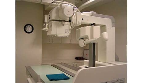
A study in the Journal of the American Academy of Orthopaedic Surgeons looks at x-Ray and MRI when it comes to evaluating knee pain among middle-age patients.
The study’s authors suggest that while MRI may help physicians diagnose problems that may be causing the knee pain, an x-ray is the best first line screening tool.
“If an X-ray shows that a person has significant arthritis, the MRI findings—like a meniscus tear—are less important because the amount of arthritis often dictates the treatment. Therefore, patients should always get a standing X-ray before getting an MRI to screen for knee pain in patients older than 40,” says lead author Muyibat Adelani, MD, an orthopaedic surgeon with Washington University’s Department of Orthopedics, in a media release from the American Academy of Orthopaedic Surgeons.
In the study, per the release, the authors looked at 100 knee MRIs from patients ages 40 and older and observed the following:
The most common diagnoses are osteoarthritis (39%), and meniscal tears (29%); Nearly one in four MRIs were taken prior to the patient’s first having obtained a weight-bearing X-ray; and, Only half of those MRIs obtained prior to meeting with an orthopaedic surgeon actually contributed to a patient’s diagnosis and treatment for osteoarthritis.
“Patients should always get weight-bearing X-rays before getting an MRI because MRIs are not always needed to diagnose knee problems,” Adelani states.
[Source(s): American Academy of Orthopaedic Surgeons, PR Newswire]


