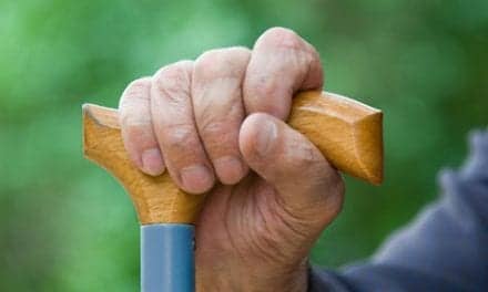
Using computed tomography (CT) to evaluate sarcopenia among elderly patients treated for fall-related injuries, researchers suggest that decreased core muscle strength could be associated with decreased survival times following a hip fracture.
The study was published recently in the American Journal of Roentgenology.
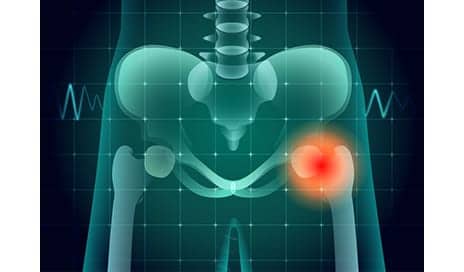
Researchers studied the records of nearly 300 people (65 years of age or older) who were treated for fall-related injuries at UC Davis Medical Center between 2000 and 2015. All patients were suspected of breaking their hips and received CT scans to diagnose or rule it out.
They then evaluated the CTs with additional measurements of the size and density of lumbar and thoracic muscle alongside the spine. That information was then compared with mortality data from the National Death Index, a centralized database of death record information maintained by the US Centers for Disease Control and Prevention.
The results showed that patients with better core muscle had significantly better survival rates over the duration of the 10-year study, explains a media release from University of California – Davis Health System.
“The fact that we were able to predict survival in such a small group of non-cancer patients is truly remarkable,” says senior author Leon Lenchik, professor of radiology at Wake Forest, in the release.
“Using CT scans to evaluate muscles in addition to hip bones can help predict longevity and personalize treatment to a patient’s needs,” states lead author Robert Boutin, a UC Davis professor of radiology. “Recognizing sarcopenia as a distinct condition that provides clues to future health can open doors to new discoveries in diagnosis and treatment.”
[Source(s): University of California – Davis Health System, Science Daily]