By pairing vital tools with prehabilitation, therapists can help enhance functional capacity prior to orthopedic surgery
Physical conditioning in preparation for surgery is termed prehabilitation1 and encompasses strength training,2 breathing exercises,3,4 and whole-body aerobic exercise training.1 While different in every individual’s practice, prehabilitation techniques have a common goal in accelerating postoperative recovery by improving physiological reserve through physical intervention.
Components of a prehabilitation exercise regimen for joint or intracavity surgery include inflammation reduction, restoration of range of motion, restoration of muscle strength, and improving aerobic capacity.
Risks of Inflammation
The inflammatory response is the body’s natural response that occurs immediately following tissue damage. Its main functions are to defend the body against harmful substances, dispose of dead or dying tissue, and promote the renewal of normal tissue. The inflammatory reaction is normally characterized by five distinct signs, each of which is due to a physiological response to tissue injury.
• Pain (due to chemicals released by damaged cells);
• Swelling or edema (due to an influx of fluid into the damaged region);
• Redness (due to vasodilatation – the widening of blood vessels and bleeding in the joint or structure);
• Heat (due to an increase in blood flow to the area); and
• Loss of function (due to increased swelling and pain).
Pain is not always bad. Pain tells us to pamper a wounded area that is trying to heal. The pain of inflammation is caused by tissue swelling that presses on nerves, as well as by chemical substances that convert a molecular signal into an electrical impulse that spells pain for the body. Acute inflammation occurs immediately after an injury and is usually short-lived. The acute inflammatory response requires constant stimulation to remain active. So, when the injury begins to heal or the source of infection has been neutralized, the symptoms of inflammation also go away.
Chronic inflammation is an unhealthy inflammatory over-response that can linger for months to years. The following is the proposed sequence of treatment which can be used to reduce this inflammatory load in the tissues with chronic inflammation. Photo-biostimulation is applied first, coincidentally with subthermal ultrasound, then is followed up with electrical stimulation to utilize the muscle pump. Finally, Kinesio tape can be used to wick away inflammation.
Photo-Biostimulation
Photo-biostimulation (laser or superluminous diodes) is light energy that penetrates the skin and is absorbed by all cell types (muscle, joint, blood, nerve, etc). Light energy is transformed into biochemical energy (similar to photosynthesis), providing additional energy. Increased ATP production and cellular cascade of events result in enhanced healing, pain relief, and restoration of normal cell function.5 Transfer gates in the cell membrane are opened, allowing rebalance of intracellular/extracellular osmotic pressure and drainage of excess intracellular fluids into the lymphatic system.
Use of photo-biostimulation over these terminal sites would open up the lymphatic flow to decrease inflammation from distal to proximal. The ease of application of Chattanooga, Tenn-based Richmar’s plug-and-play MedX 785-nm laser with 200 mW Gallium-Aluminum-Arsenide diodes on the EVO CM4 provides quick treatment of the terminal sites at 4 J/cm2 or 20 seconds (the laser generates 1 joule every 5 seconds) every other day, or if you utilized the plug-and-play superluminous diodes (SLD), you could coincidentally treat the terminal sites while applying ultrasound on the same Richmar EVO CM4 to reduce inflammation at the local site. The SLD infrared (870 nm) and the visible red (633 nm) provide similar outcomes to the laser to open up those terminal sites, but takes a little longer at 1 Joule per 45 seconds application (or a 3-minute treatment) to be applied every other day.
Richmar’s EVO CM4 ultrasound cues up the subthermal application with a single button when applied. Coincidentally with photo-biostimulation, it will utilize the 3 MHz sound head at 0.5 w/cm2 x 5 minutes over the actual inflamed or the now edematous region, which will change cell membrane permeability by a process of acoustic streaming and stable cavitation. Ultrasound will also modulate vasoconstriction; change lymphocyte adhesion properties of the endothelium; increase mast cell degranulation; increase phagocytosis by macrophage; increase production of growth factors by macrophages, resulting in calcium fluxes in fibroblasts, angiogenesis, proliferation of T-cells, osteoblasts, fibroblasts, and a number of proteins associated with inflammation and repair (IL-1, IL-2, IL-6, IL-8, interferon-?, fibroblast growth factor-b, vascular endothelial growth factor, collagen),6,7,8-10,11-19; as well as accelerate thrombolysis20-29 or reduce inflammation. The Accusound applicator is an option with the Richmar EVO CM4 that aims to make ultrasound application hands-free, and eliminate all the associated faux pas (Draper’s top 10 faux pas).
One source for ultrasound applicator gel is Parker Laboratories Inc, Fairfield, NJ, which offers product lines including Aquasonic 100 ultrasound transmission gel and Aquasonic Clear ultrasound gel.
In terms of low-level-lasers, additional technologies available include the Vectra Genisys therapy system (Chattanooga, a DJO Global Company, Vista, Calif) and Apollo Class IV cold lasers from PHS Therapeutics, a division of Pivotal Health Solutions, Watertown, SD.
Pain as a Limiting Factor
Repeated pain exposure bombards pain synapses with repetitive input, increasing their responsiveness to later stimuli, through a process similar to learning. Therefore, although the individual may learn cognitive methods of coping with pain, these methods may not be sufficient to cope with the boosted response to future painful stimuli (like surgery).30 Kalat suggests that morphine should be taken before surgery.30 “People who begin taking morphine before surgery need less of it afterward.”31
New research has suggested that preventing the nervous system from being overtaxed by pain from the trauma of surgery may lead to a less painful postoperative experience. Pretreated patients may require less postsurgical medications, and they may recover more quickly, possibly experiencing pain-free days far sooner than patients who have used traditional postsurgical pain methods. The concept of pain management would be to elevate or maintain the surgical candidate’s pain threshold. Pain as a limiting factor in prehabilitation can be addressed with photo-biostimulation, ultrasound, electrical stimulation and therapeutic tape, and body weight-supported gait training.
Photo-biostimulation at dosages for acute pain 4 J/cm2 and for chronic pain 10 J/cm2 are recommended. Scott D. Fender and David Diffee of the Pain Research Group in Arvada, Colo, suggest targeting the area of the stellate ganglion in the rehabilitation of patients with a history of chronic musculoskeletal pain syndromes. Application of photo-biostimulation over acupuncture points in the arm at LI4, LI1, Tb5, and P6 are also effective means of managing pain anywhere in the body. Pain relief from ultrasound treatment occurs through transmitter signal substances and direct effect on nerves with an increase in endorphins, serotonin, acetylcholine, nitric oxide,32 and through the peripheral and central nerve by a decrease in C fiber activity by blocking depolarization and increased replenishment of neurotransmitters at the synaptic level.33 The same parameters and rationale for subthermal ultrasound for inflammation management will be used to reduce pain.
An easy method for pain management would be the application of electrical stimulation. A single preset button on the Richmar EVO CM4 offers the Quadpolar or Premodulated Interferential current (chained 100 Hz x 15 minutes and 2 Hz x 15 minutes, vector on fast 90o with an intensity at a slight motor twitch) or Microcurrent for the e-stim shy patient (chained 30 Hz x 15 minutes and 0.3 Hz x 15 minutes with a subsensory intensity). Electrodes are applied to the same four acupuncture points at LI4, LI11, Tb5, and P6 to affect Melzack and Wall’s Gate Control Theory or Bausbaum and Fields’ endogenous opiate theory. A portable TENS unit would be a means of carrying over the positive pain-management results that you would incur in the clinic.34
A wide variety of electromedical modalities can be found on the market. Several of these device models are designed to provide multiple types of treatment from a single unit. These include the UltraTENSII Ultrasound and TENS combination unit from Roscoe Medical, Strongsville, Ohio. To manage acute and chronic pain, Amrex, a division of Amrex-Zetron Inc, Paramount, Calif, offers the SpectrumMicro-1000, which provides eased treatment protocol setup, a selectable audio function indicator, and “quick read” illuminated visual function indicators.
An alternative to manage inflammation and pain would be iontophoresis or a transdermal delivery system. This method can be used to drive a drug across the skin barrier. Examples of iontophoresis-based products include the ActivaPatch and Trivarion Butterfly, available through ActivaTek Inc, Gilroy, Calif, and the IontoPatch from Patterson Medical, headquartered in Warrenville, Ill.
In any healthy joint, the bones, ligaments, tendons, cartilaginous structures, and muscles help to stabilize the joint. These may all be compromised, and the muscle and tendon strength will be starting to deteriorate due to pain and lack of use due to pathology. Therefore, maintaining or gaining strength in the muscles that support the joint in prehabilitation is required to help support the stability of that joint.
During voluntary contraction, motor units are recruited asynchronously and according to the size principle, in order from slow, type-I motor units to fast, type-II motor units.35 During electrically evoked muscle contraction, motor units fire synchronously and in reverse order from fast, type-II motor units to slow type-I motor units.36,37 The combination of two physiological processes—ie, the reverse order of motor unit recruitment combined with external loading (muscle hypertrophy) imposed on stimulated muscles during training—are likely responsible for the gains in muscular strength observed during muscle re-education training. Studies suggest that e-stim for muscle re-education produces lasting gains in muscle force through a short-term training regimen (approximately 30 training sessions).38-42
Altered gait biomechanics is common amount individuals with hip, knee, or ankle pathologies, especially those with rheumatoid arthritis and osteoarthritis (OA). Reduced motion of the knee joint leads to increased pelvic motion, which should affect the natural mobility of the lumbar spine and cause pain in the lumbar region of the spine due to kinematic interaction.43 People with joint and neurological deficits need to learn to move again in the most energy-efficient way possible within the confines of the damage sustained. The therapist is therefore a mediator in each individual’s motor learning process, especially in prehabilitation.
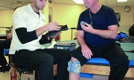
The key takeaway from motor learning is that practicing skills over time causes those neural pathways to work better in unison via myelination. Practice that is performed frequently and correctly in addition to useful feedback are necessary in improving performance. LiteGait, available through Mobility Research, Tempe, Ariz, is a postural control device that provides posture, balance, and weight-bearing assistance while correcting upright postures appropriate for ambulation both over a treadmill and over ground.
LiteGait enables a partial weight-bearing environment to allow the therapist the freedom to facilitate weight shifting to correct an antalgic gait with hip problems or off-loading sufficiently the lower extremity, as in a painful OA knee, to facilitate foot placement or to cue proper patterns of walking. LiteGait can distribute supporting forces to meet patient needs to become more symmetric in static posture and more functional in dynamic gait. LiteGait is also designed to help increase the stretch of the hip flexors to facilitate the central pattern generators (automatic gait) or to integrate knee extension to increase pain-free weight-bearing in midstance.
Additional body weight support systems include the Lokomat Pro, available through Hocoma, NorWell, Mass, which can be used to control weight bearing in a harness-over-treadmill design; the Biodex Unweighing System, Biodex Medical Systems Inc, Shirley, NY; and the Solo-Step System, from Sioux Falls, SD-based Solo-Step.
Topicals and Hot/Cold Therapy
Topical analgesics, such as the pain-relieving gels marketed by Sombra, headquartered in Albuquerque, NM, Ari-Med Pharmaceuticals, Tempe, Ariz, and Sore No More, based in Moab, Utah, can also be used to target relief from conditions that include joint and muscle pain.
Heat therapy can allow for the dilation of blood vessels and improvement of oxygen flow and circulation to promote healing. Added control of pain can be offered through cold therapy, which can help restrict the blood vessels to reduce swelling or numb injured tissue by slowing the nerve impulse. Prehabilitation improves an individual’s functional capacity through increased physical activity before an anticipated orthopedic procedure (surgery). If an individual maintains a higher level of functional ability before a procedure, they will rebound more rapidly in rehabilitation postprocedure. Prehabilitation will enhance an individual’s functional capacity to withstand the stressor of inactivity associated with an orthopedic procedure by managing inflammation, pain, muscle strength, range of motion, and gait deviations, which are all accomplished with the tools that are already present in our physical therapy toolbox. PTP
Keith Khoo, PT, graduated from the University of Oklahoma and has been in practice since 1985. At the Scholl Center, Tulsa, Okla, he evaluates and treats movement disorders such as Parkinson’s disease and multiple sclerosis, as well as neuropathic patients, balance and vestibular disorders, pain management, and headache patients. Khoo has taught continuing education courses on modality applications for neuromusculoskeletal dysfunctions, vestibular and balance dysfunction, and Kinesio taping in the United States and abroad. For more information, contact [email protected].
References
1. Carli F, Zavorsky GS. Optimising functional exercise capacity in theelderly surgical population. Curr Opin Clin Nutr Metab Care. 2005;8:23-32.
2. Swank AM, Kachelman JB, Bibeau W, et al. Prehabilitation before total knee arthroplasty increases strength and function in older adults with severe osteoarthritis. J Strength Cond Res. 2007; 25:318-325.
3. Dronkers J, Veldman A, Hoberg E, van derWaal C, van Meeteren N. Prevention of pulmonary complications after upper abdominal surgery by preoperative intensive inspiratory muscle training: a randomized controlled pilot study. Clin Rehabil. 2008;22:134-142.
4. Hulzebos EH, Helders PJ, Favié NJ, De Bie RA, Brutel de la Riviere A, Van Meeteren NL. Preoperative intensive inspiratory muscle training to prevent postoperative pulmonary complications in high risk patients undergoing CABG surgery: a randomized clinical trial. J Am Med Assoc. 2006; 296: 1851-1857.
5. Tuner J, Jode L. The Laser Therapy Handbook. Prima Books; 2004.
6. Dyson M. Mechanisms involved in therapeutic ultrasound. Physiotherapy. 1987;73:116-120.
7. Love LA, Kremkau FW. Intracellular temperature distribution produced by ultrasound. J Accoust Soc Am. 1980;67:1045-1050.
8. Fischell TA, Abbas MA, Grant GW, Siegel RJ. Ultrasonic energy: effects on vascular function and integrity. Circulation. 1991;84:1783-1795.
9. Maxwell L, Collecutt T, Gledhill M, Sharma S, Edgar S, Gavin JB. The augmentation of leucocytes adhesion to endothelium by therapeutic ultrasound. Ultrasound Med Biol. 1994;20:383-390.
10. Steffen W, Cumberland D, Gaines P, et al. Catheter-delivered high intensity, low frequency ultrasound induces vasodilation in vivo. Eur Heart J. 1994;15:369-376.
11. Doan N, Reher P, Meghji S, Harris M. In vitro effects of therapeutic ultrasound on cell proliferation, protein synthesis and cytokine production by human fibroblasts, osteoblasts and monocytes. J Oral Maxillofac Surg. 1999;57:409-419.
12. Dyson M, Luke D A. Induction of mast cell degranulation in skin by ultrasound. IEEE Trans Ultrason Ferroelectr Freq Control. 1986;33:194-201.
13. Hogan RD, Franklin TD, Fry F , Avery KA, Burke KM. The effect of ultrasound on microvascular hemodynamics in skeletal muscle: effect on arterioles. Ultrasound Med Biol.1982;8:45-55.
14. Johns LD, Colloton PA, Neuenfeldt J. Effects of therapeutic ultrasound on T cell proliferation and IL-2 production [abstract]. J Athl Train. 1999;34(suppl):S24.
15. Mortimer AJ, Dyson M. The effect of therapeutic ultrasound on calcium uptake in fibroblasts. Ultrasound Med Biol. 1988;14:499-506.
16. Reher P, Doan N, Bradnock B, Meghji S, Harris M. Effect of ultrasound on the production of IL-8, basic FGF and VEGF. Cytokine. 1999;11:416-423.
17. Vivino AA, Boraker DK, Miller D, Nyborg W. Stable cavitation at low ultrasonic intensities induces cell death and inhibits 3H-TdR incorporation by Con-A-stimulated murine lymphocytes in vitro. Ultrasound Med Biol. 1985;11:751-759.
18. Young SR, Dyson M. The effect of therapeutic ultrasound on angiogenesis. Ultrasound Med Biol.1989;16:261-269.
19. Young SR, Dyson M. Macrophage responsiveness to therapeutic ultrasound. Ultrasound Med Biol.1990;16:809-816.
20. Blinc A, Francis CW, Trudnowski JL, Carstensen E. Characterization of ultrasound-potentiated fibrolysis in vitro. Blood. 1993;81:2636-2643.
21. Francis CW, Onundarson PT, Carstensen EL, et al. Enhancement of fibinolysis in vitro by ultrasound. J Clin Invest. 1992;90:2063-2068.
22. Harpaz D, Chen X, Francis CW, Marder VJ, Metzer RS. Ultrasound enhancement of thrombolysis and reperfusion in vitro. J Am Coll Cardiol. 1993;21:1507-1511.
23. Higazi AA, Katz I, Mayer M, Weiss AT. The effect of ultrasonic irradiation and temperature on fibrinolytic activity in vitro. Thromb Res. 1993;69:251-253.
24. Kornowski R, Meltzer RS, Chernine A, Vered Z, Battler A. Does external ultrasound accelerate thrombolysis? Results from a rabbit model. Circulation. 1994;89:339-344.
25. Kudo S. Thrombolysis with ultrasound effect. Tokyo Jikeikai Med J. 1989;104:1005-1012.
26. Lauer CG, Burge R, Tang DB, Bass BG, Gomez ER, Alving BM. Effect of ultrasound on tissue-type plasminogen activator-induced thrombolysis. Circulation. 1992;86:1257-1264.
27. Luo H, Steffen W, Cercek B, Arunasalam S, Maurer G, Siegel RJ. Enhancement of thrombolysis by external ultrasound. Am Heart J. 1993;125:1564-1569.
28. Nilsson AM, Odselius R, Roijer A, Olsson SB. Pro- and antifibrinolytic effects of ultrasound on streptokinase-induced thrombolysis. Ultrasound Med Biol. 1995;21:833-840.
29. Olsson SB, Johansson B, Nilsson AM, Olsson C, Roijer A. Enhancement of thrombolysis by ultrasound. Ultrasound Med Biol. 1994;20:375-382.
30. Kalat JW. Biological Psychology. 9th ed. Belmont, Calif: Thomson/Wadworth; 2007:212
31. Keefe FJ, France CR. Pain: biopsychosocial mechanisms and management. Curr Dir Psychol Sci. 1999;8(5):137.
32. Bjordal JM, Johnson M, Iversen V, Aimbire F, Lopes-martins, RAB. Low-level laser therapy in acute pain: a systematic review of possible mechanisms of action and clinical effects in randomized placebo-controlled trials. Photomed Laser Surg. 2006;24(2):237-247.
33. Basford JR, Hallman HO, Matsumoto JY, Moyer SK, Buss JM, Baxter GD. Effects of 830nm continues wave laser diode irradiation on median nerve function in normal subjects. Lasers Surg Med. 1993;13(6):597-604.
34. Cheing GL, Hui-Chan CW. Analgesic effects of transcutaneous electrical nerve stimulation and interferential currents on heat pain in healthy subjects. J Rehab Med. 2003;35(1):15-19.
35. Henneman E, Olson CB. Relations between structure and function in the design of skeletal muscles. J Neurophysiol. 1965;28:581-598.
36. Kraemer WJ, Deschenes MR, Fleck SJ. Physiological adaptations to resistance exercise. Implications for athletic conditioning. Sports Med. 1988;6(4):246-256.
37. Delitto A, Brown M, Strube MJ, Rose SJ, Lehman RC. Electrical stimulation of the quadriceps femoris in an elite weight lifter. J Sports Med. 1989;10(3):187-191.
38. Selkowitz DM. Improvement in isometric strength of the quadriceps femoris muscle after training with electrical stimulation. Phys Ther. 1985;65(2):186-196.
39. Laughman RK, Youdas, JW, Garrett TR, Chao EY. Strength changes in normal quadriceps femoris muscle as a result of electrical stimulation. Phys Ther. 1983;63(4):494-499.
40. Soo CL,Currier DP, Threlkeld AJ. Augmenting voluntary torque of healthy muscle by optimization of electrical stimulation. Phys Ther. 1988;68(3):333-337.
41. Kubiak RJ, Whitman KM, Johnston RM. Changes in quadriceps femoris muscle strength using isometric exercise versus electrical stimulation. J Orthop Sports Phys Ther. 1987;8(11):537-541.
42. Currier DP, Mann R. Muscular strength development by electrical stimulation in healthy individuals. Phys Ther. 1983;63(6):915-921.
43. Bejek Z, Paróczai R, Illyes A, Kocsis L, Kiss RM. Gait parameters of patients with osteoarthritis of the knee joint. Phys Educ Sport. 2006;4(1):9-16.